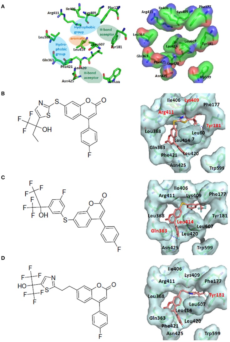
Figure 8.
LeadOp+R result for the 5-LOX kinase model system. (A) Schematic representation of the human 5-LOX active site (left) and the binding pocket (right). The purported pharmacophores of the binding site of 5-LOX involving two hydrophobic groups (blue ovals), two hydrogen bond acceptors (green ovals), and an aromatic ring (orange oval) for ligand binding at the binding cavity. (B–D) Chemical structure (left) and MDS result (right) of the generated compound rB1 (B), the generated compound rB2 (C), and the generated compound rB3 (D). Carbon atoms are colored pink. Amino acid residues that participate in hydrogen-bonding interactions (labeled red) with the proposed compound within the binding site are depicted with gray molecular surfaces.

