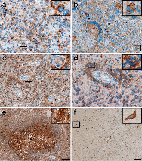
Fig. 2.

Immunohistochemical expression of CD63 in glioblastoma
a A distinct cytoplasmic CD63 staining was seen in all tumors (arrow). b A distinct plasma membrane labeling was seen in some biopsies (arrows). c, d In general blood vessels showed a weak CD63 expression, but a more intense expression was found in perivascular tumor cells (d). e Pseudopalisade formations showed an intense CD63 expression. f A few CD63 tumor cells with faint CD63 expression as well as CD63+ pyramidal shaped neurons (arrow) were seen in the tumor periphery. Scale bar: 50 μm (a-d) and 100 μm (e-f)
