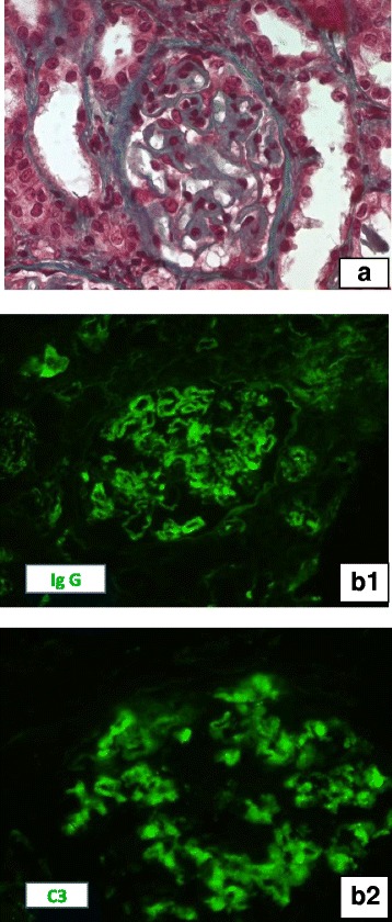
Fig. 1.

Kidney biopsy Light microscopy (A): A glomerulus with important thickening of glomerular capillary wall and normal cellularity(Masson Trichrom, × 400). Immunofluorescence studies: Intense granular staining of IgG (B1) and C3 (B2) along the glomerular capillaries (direct immunofluorescence, × 400)
