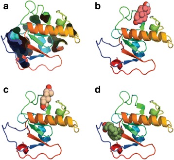
Fig. 5.

The hydrophilic segment model of PGRMC. The structure is represented as cartoons with N-terminus in blue and the C-terminus in red. In a the cavities are represented as surfaces in the progesterone docking, b shows docking of testosterone, c dehydroepiandrosterone, and d arachidonic acid, all of them in a CPK representation. Note that both dehydroepiandrosterone and arachidonic acid bind to a similar site whereas progesterone binds closer to the C-terminus
