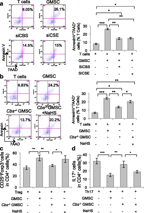
Fig. 2.

H2S was required for GMSCs to induce T-cell apoptosis. a In GMSC and T-cell coculture, GMSCs induced T-cell apoptosis, while CBS or CSE siRNA treatment attenuated GMSC-induced T-cell apoptosis, as assessed by flow cytometry (n = 5). b Cbs−/− GMSCs induced less T-cell apoptosis compared with control GMSCs, and treatment with NaHS elevated T-cell apoptosis induced by Cbs−/− GMSCs (n = 5). c GMSCs promoted T-cell differentiation into Treg cells induced in their polarized condition. Cbs−/− GMSCs showed decreased capacity to promote Treg cell differentiation, while NaHS treatment partially restored Treg cell polarization induced by Cbs−/− GMSCs (n = 5), as assessed by flow cytometry. d GMSCs inhibited T-cell differentiation into Th17 cells in a Th17 cell polarization condition. Cbs−/− GMSCs showed a decreased capacity to inhibit Th17 cell differentiation, while NaHS treatment partially restored the capacity of inhibition of Th17 cell polarization by Cbs−/− GMSCs, as assessed by flow cytometry (n = 5). *P < 0.05, **P < 0.01, ***P < 0.001. Error bars: mean ± SD. All experimental data verified in at least three independent experiments. CBS cystathionine β-synthase, CSE cystathionine γ-lyase, GMSC gingiva-derived mesenchymal stem cell, si small interfering
