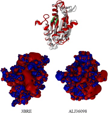
Fig. 3.

Structure of ALJ36098 from the A. brasilense Sp7 genome. a The model superimposed with WspR (PDB id: 3BRE) from P. aeruginosa [47]. The 3BRE structure is colored red. The GGDEF motif is colored green, and the SDHAF motif is shown superimposed. b The electrostatic potential (+/˗ 4kT/e) calculated with the APBS of both proteins in the same orientation, as indicated above (a). The positive amino acid residues are colored blue, and the negative amino acid residues are colored red. The potential difference in the binding site changes in ALJ36098
