Fig. 2.

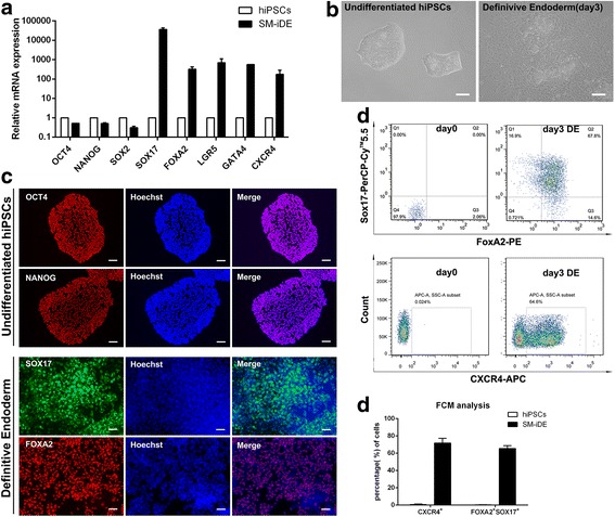
Small molecules efficiently induce definitive endoderm differentiation from hPSCs. a qRT-PCR for pluripotent markers and DE markers using RNA lysates from human iPSCs treated with DMSO at day 1, CHIR99021 (3 μM) at day 2, and then basal medium without CHIR99021 at day 3. b Phase contrast photos (×200) showing morphological changes during stage I of differentiation. Scale bars = 100 μm. c Immunofluorescence of pluripotency and DE-specific markers at the end of differentiation stage I. Scale bars = 100 μm. d, e Percentage of FOXA2/SOX17- and CXCR4-positive cells at day 0 and day 3 of DE differentiation analyzed by flow cytometry. e Histogram of the FOXA2/SOX17- and CXCR4-positive cells at day 0 and day 3 of DE differentiation analyzed by flow cytometry. Undifferentiated human iPSCs were used as control. All data were presented as the mean of at least three independent experiments. The error bars represent the SD
