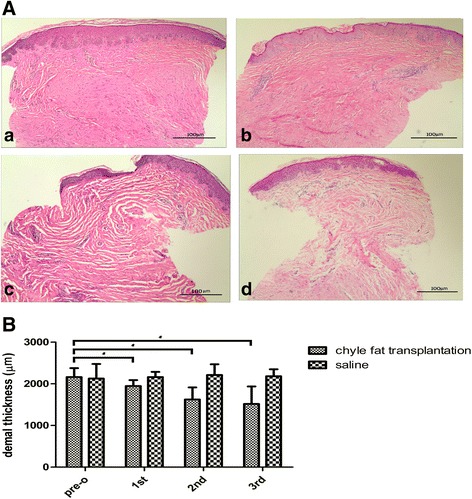
Fig. 4.

Histopathologic evaluation of a hypertrophic scar. A Compared with the preoperative scar (a), after three treatments the dermis was thinner than before (b–d), density and quantity of fibroblasts were decreased, and density of blood vessels was reduced. Each patient was treated three times, with interval between treatments of 3 months. Cells stained with hematoxylin and eosin (magnification × 100). B Chyle fat transplantation treatment gradually improved the dermal thickness of hypertrophic scars.
*pre-o chyle fat transplantation group compared with 1st, 2nd and 3rd chyle fat transplantation group , P < 0.05
