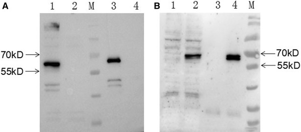
Figure 3.

Immunoprecipitation analysis. LMH cells were infected with FAdV-4 or transfected with pcDNA3.1-F2. Then, 3C2 was used as the capture antibody to perform immunoprecipitation, followed by Western blot analysis using chicken sera against FAdV-4. A Lanes 1 and 2: lysates of LMH cells infected with or without FAdV-4, respectively. Lanes 3 and 4: 3C2-immunoprecipitated pellets of LMH cells infected with or without FAdV-4, respectively. B Lanes 1 and 2: lysates of LMH cells transfected without or with pcDNA3.1-F2, respectively. Lanes 3 and 4: 3C2-immunoprecipitated pellets of LMH cells transfected without or with pcDNA3.1-F2, respectively.
