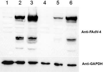
Figure 5.

Western blot analysis of the neutralization activity of 3C2. 1:200 and 1:5000 dilutions of 3C2 or control 6E6 mAbs were first mixed with FAdV-4, and then LMH cells were infected with the mixture and analysed by Western blot using chicken sera against FAdV-4, as described in “Materials and methods”. Lanes 1 and 2: lysates of LMH cells infected with the mixture of virus and the 1:200 dilutions of 3C2 and 6E6, respectively. Lanes 3 and 4: lysates of LMH cells infected with or without FAdV-4, respectively. Lanes 5 and 6: lysates of LMH cells infected with the mixture of virus and 1:5000 dilutions of 3C2 and 6E6, respectively.
