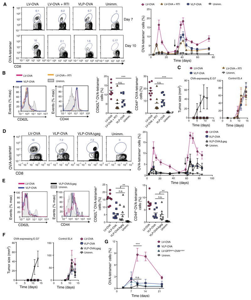
Fig. 3. LV DNA, genome, and capsid are not required for DC activation and CD8+ T cell priming in vivo.
(A) Wild-type mice received homologous prime-boost vaccination of SVGmu-pseudotyped LV-OVA with or without RTI or VLP-OVA (n = 8 mice per group). Representative FACS plots show OVA-tetramer+ cells gated on CD8+ T cells from the blood at 7 and 10 days after primary immunization (left). Graph depicts percentages of OVA-tetramer+ CD8+ T cells from the blood of immunized and unimmunized mice over time (black arrow, boost) (right). (B) Representative FACS plots show expression of CD62L and CD44 on OVA- tetramer+ CD8+ T cells from immunized mice compared with naïve CD8+ T cells from unimmunized mice at 7 days after boost (left). Graphs depict percentages of CD62Llo and CD44hi OVA-tetramer+ cells, with each symbol representing an individual mouse and horizontal bar indicating the mean (right). (C) Seven weeks after boost, mice were injected with 5 × 10⁶ OVA-expressing E.G7 thymoma tumor cells and 5 × 10⁶ EL4 (control) non–OVA-expressing EL4 thymoma tumor cells on opposing legs, and tumor sizes were measured. (D to F) Wild-type mice were homologously prime-boosted with LV-OVA, VLP-OVA, or capsid-less VLP-OVAΔgag. OVA-tetramer+ cells from the blood were analyzed as in (A) (n = 8 mice per group) at 7 days after boost (left) and over time (right) (D). CD62L and CD44 expression of OVA-tetramer+ CD8+ T cells from immunized mice compared with naïve CD8+ T cells from unimmunized mice at 7 days after boost were measured as in (B) (E). Mice were injected with tumor cells, as in (C), and tumor sizes were measured (F). (G) Wild-type mice were immunized with LV-OVA, LV encoding OVA carrying GFP (LV-GFPgene-OVAprotein), or VLP-OVA (n = 8 mice per group), and OVA-tetramer+ CD8+ T cells from the blood were measured over time. Statistical comparisons were made between the LV-GFPgene-OVAprotein– and VLP-OVA– or LV-OVA–immunized mice. Data are representative of two independent experiments (A to G). Results are shown as mean ± SEM (A, C, D, F, and G). P > 0.05; **P < 0.005; ***P < 0.001 (unpaired Student’s t test).

