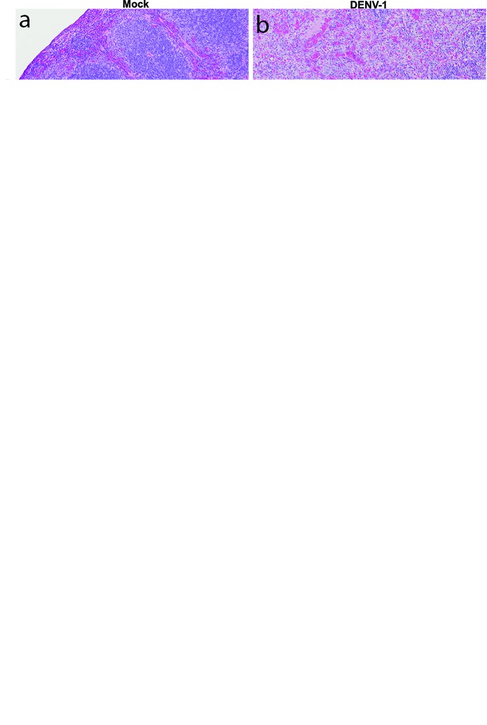
Fig. 5.
Histological changes resulting from DENV-1 WP 74 infection; H&E section micrograph images from AG129 mice mock-infected (a, c, e, g) or infected with 7.4 log10 p.f.u. DENV-1 WP 74 (b, d, f, h) show the major changes seen in infected animals. (a, b) Ultrastructural changes in the virus infected spleen, with breakdown of normal architecture in the DENV-1-infected section. (c, d) Higher magnification images showing activated cells throughout the infected section. (e, f) Normal liver architecture in the control sections, but inflammation and focal necrosis in the DENV-1-infected section. (g, h) Areas of cellular expansion outside the Peyer’s patches in the large intestine of DENV-1-infected animals. Magnification: ×4 (a, b, e, f, g, h) or ×10 (c, d)

