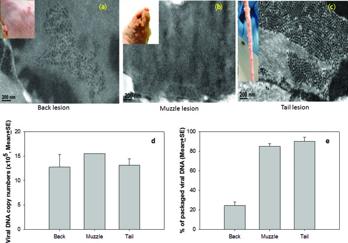
Fig. 6.
Viral particle detection by transmission electron microscopy (TEM). From many fields of view, only a few infected cells, each containing relatively few viral particles, were identified in the back (a), while the muzzle (b) and the tail (c) lesions showed abundant infected cells filled with viral particles. No significant difference was found in the viral DNA copy numbers between the tail, the muzzle and the back lesions by Q-PCR analysis (d, P>0.05, one way ANOVA analysis). However, viral DNA packaging rates were significantly lower in the back lesions when compared to those in the muzzle and tail lesions (e, P<0.01, one way ANOVA analysis).

