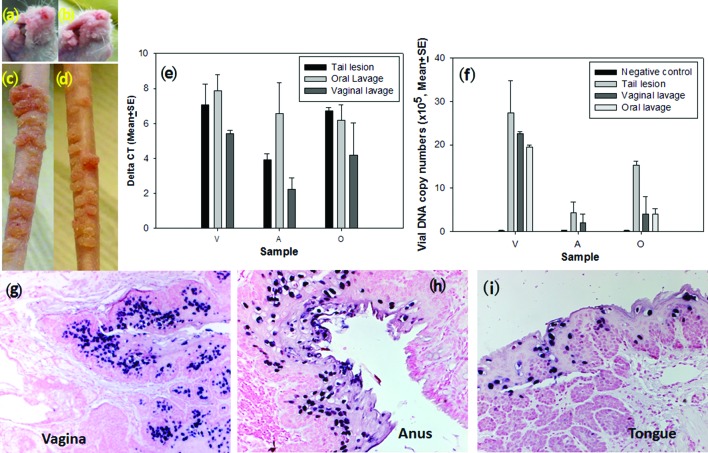
Fig. 7.
Viral infection with vaginal or oral lavage samples. Representative muzzle (a, b) and tail (c, d) lesions induced by either vaginal lavage extracts (a, c) or oral lavage extract (b, d) in the outbred nude mouse at 8.5 months post-infection. The lesions became evident around week three post-infection and continued to grow over the period of observation. The lesions were comparable to those induced by the virus harvested from the tail lesions used in our previous studies. (e) Detection of viral DNA by SYBR Green Q-PCR analysis from mucosal [vaginal (V), anal (A) and oral (O)] sites infected with viral suspension from tail lesion (control), oral or vaginal lavages. Comparable viral DNA copy numbers were found in all three mucosal sites infected from the three sources of viruses (e and f, P>0.05, one-way ANOVA analysis). Lavage samples from naïve mice without MmuPV1 infection were used as the negative control. Viral DNA was detected at the vaginal (g), anal (h) and oral (i) infected sites by in situ hybridization.

