
Fig. 1.
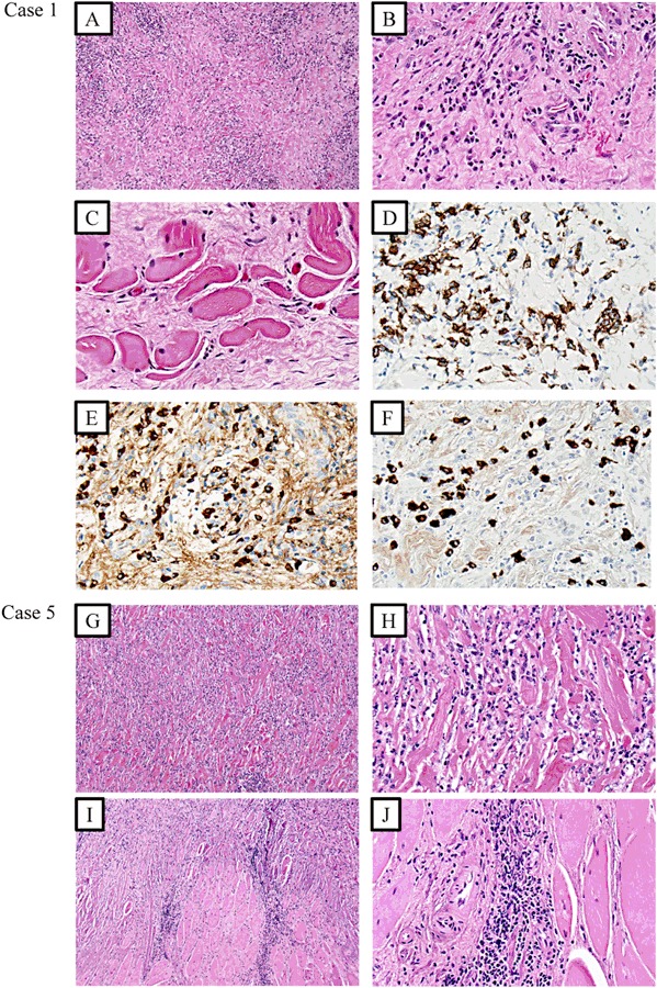
Photomicrographs of case 1 (a–f) and case 5 (g–j) with representative features of Riedel´s thyroiditis. All photos except (d–f) represent haematoxylin-eosin stainings. a, b Storiform, keloid-like fibrosis, and inflammatory cells have replaced the thyroid parenchyma (magnified x100 and x400 respectively). c The inflammatory cells and fibrosis engage the perithyroidal skeletal musculature (x400 magnification). d CD138 immunohistochemistry visualizes the infiltrative plasma cells (x400 magnification). e, f IgG-positive and IgG4-positive cells respectively in the same area, cells at x400 magnification. The number of IgG4-positive cells are >10/high power field, and the IgG4/IgG ratio is >0.5, suggestive of IgG4-related disease. g, h Storiform, keloid-like fibrosis, and inflammatory cells have replaced the thyroid parenchyma (magnified x100 and x400 respectively). i, j The inflammatory cells and fibrosis engage the perithyroidal skeletal musculature (x100 and x400 magnification respectively)