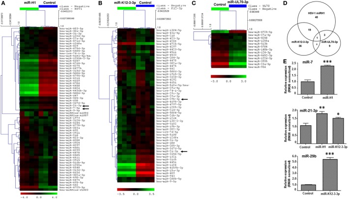
Figure 1.
v-miRs modulate expression of cellular miRNAs in human oral keratinocytes (HOK). Heat maps showing differentially expressed cellular miRNAs in (A) miR-H1, (B) miR-K12-3-3p, and (C) miR-UL70-3p transfected HOK. (D) Venn diagram showing the distribution of unique and overlapping altered cellular miRNAs in v-miR-transfected HOK. (E) Validation of microarray by quantitative RT-PCR for miR-7, miR-21-3p, and miR-29b (marked by black arrows in the heat map) in another separate cohort of transfected HOK. RNU6 was used as endogenous control. Data are expressed as mean ± SEM of four independent transfections. Student’s t-test was conducted to calculate p-values (*p < 0.05, **p < 0.01, ***p < 0.001).

