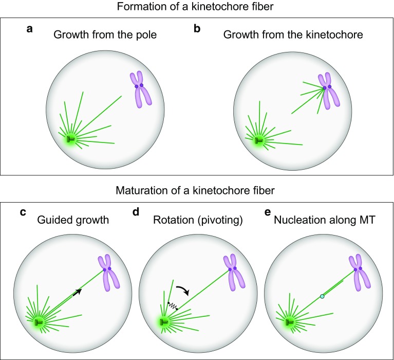
Fig. 4.
Formation (a, b) and maturation (c–e) of kinetochore fibers. a, b Microtubules of the future kinetochore fiber are formed at the spindle pole or at the kinetochore, respectively. c New microtubules are formed at the pole and grow along the existing kinetochore microtubules. d New microtubules are formed at the pole and grow at an angle with respect to the existing microtubules, forming a V-shape. They rotate around the spindle pole and eventually approach the existing kinetochore microtubules, which is followed by their binding via crosslinking proteins (black spring). e New microtubules are nucleated at the nucleation sites (light blue) along the existing kinetochore fiber. In all panels, microtubules are shown as green lines, centrosomes as green circles with small cylinders representing centrioles inside, and chromosomes are purple with kinetochores depicted as dark purple circles

