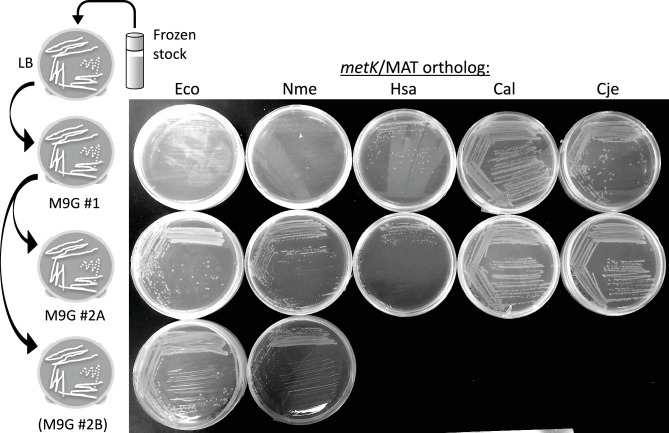
Fig. 3.
Growth of various complemented strains on minimal glucose agar plates. The experiment is shown schematically at the left, with cells taken from a frozen stock (LB broth +20 % glycerol, stored at −80 °C), streaked onto LB agar (not shown in the photograph) and then isolated colonies re-streaked onto minimal glucose agar (top row). Colonies from these glucose plates were then re-streaked onto fresh minimal glucose plates (bottom two rows). All plates were incubated for 24 h at 37 °C and then refrigerated until photographed.

