Figure 2.
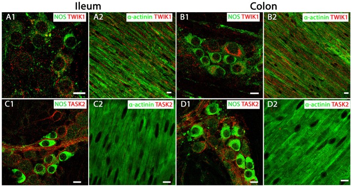
TWIK-1 and TASK-2 channels are distributed in myenteric plexus inhibitory neurons and longitudinal smooth muscle layers of the mouse ileum and colon. Panels show overlays of cell specific markers (green) and individual K2P channel markers (red) throughout. (A1,B1) show overlays for NOS immunoreactive myenteric plexus neurons and the TWIK-1 channel in ileum and colon respectively. TWIK-1 labeling was cytoplasmic and can be observed in both NOS-positive and NOS-negative enteric neurons. Within the longitudinal smooth muscle layer, TWIK-1 channels were distributed in α-actinin-immunopositive smooth muscles in both ileum (A2) and colon (B2). (C1,D1) show overlays for NOS immunoreactive myenteric plexus neurons and the TASK-2 channel in ileum and colon respectively. TASK-2 displayed cell membrane labeling of NOS-negative cell bodies and axons. Immunoreactivity for the TASK-2 channel was not detectable in α-actinin immunopositive longitudinal smooth muscle cells in either mouse ileum (C2) or colon (D2). Scale bars represent 10 μm.

