Figure 4.

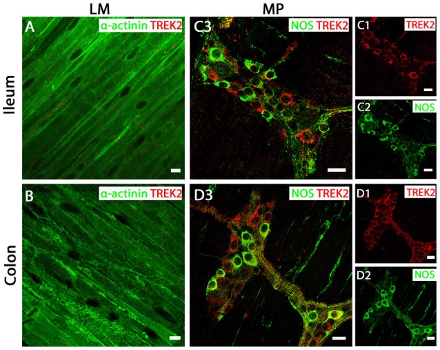
TREK-2 channels are expressed in myenteric plexus (MP) neurons of both mouse ileum and colon. (A,C3) (ileum) and (B,D3) (colon) show overlays of cell specific markers (green) and the TREK-2 channel marker (red). For clarity, (C1,C2) (ileum) and (D1,D2) (colon) show individual images from corresponding positive merges in enteric neurons. Within the longitudinal smooth muscle (LM) layer, the TREK-2 channel signal was weak and barely detectable in α-actinin immunopositive smooth muscle cells in both ileum (A) and colon (B). (C3,D3) show overlays for NOS immunoreactive myenteric plexus neurons and the TREK-2 channel in ileum and colon respectively. TREK-2 channels appear to be distributed in both NOS-positive and NOS-negative enteric neurons. Scale bars represent 10 μm (A,B) and 20 μm (C1–C3,D1–D3).
