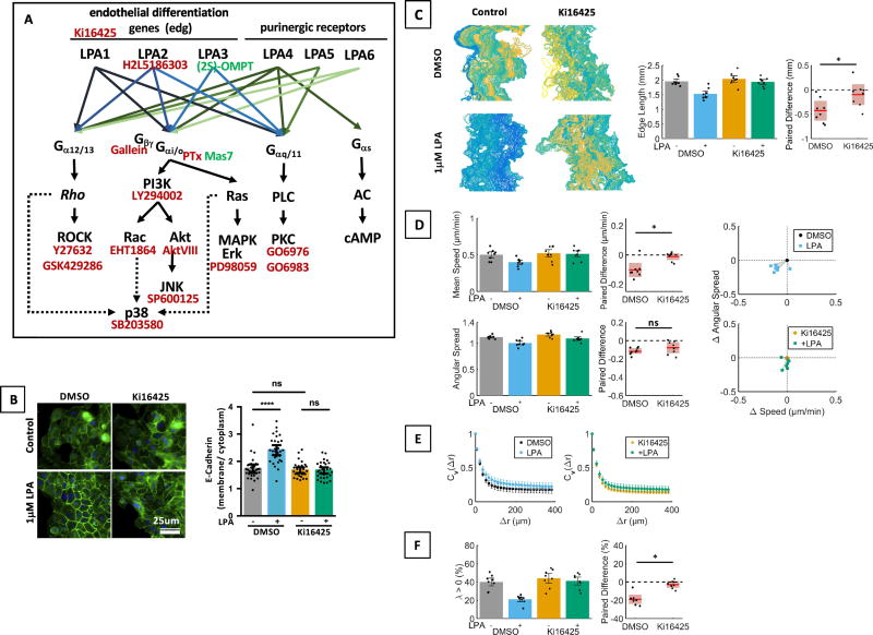
Fig. 3. Effects of LPA on MCF10CA1a cell migration are receptor mediated.
A. Schematic of the LPA signaling network (modified from [4,16]) and inhibitors used in this study. B. LPA mediated E-cadherin localization was reduced by treatment of MCF10CA1a cells with Ki16425 (10µM) as shown by immunofluor bescence. n = 30 cells per group, representative of 4 experiments. Kruskal Wallis test / Dunn’s Multiple comparison. Scale bar applies to all images of this panel. C–F. Inhibition of LPA1,3 with Ki16425 reduced the effect of LPA on edge length (C, paired t-test), cell speed (Wilcoxon matched-pairs sign rank test) and angular spread (albeit not significantly, paired t-test, D), velocity autocorrelations (E), and λ (Wilcoxon matched-pairs sign rank test, F). n = 8 experiments.

