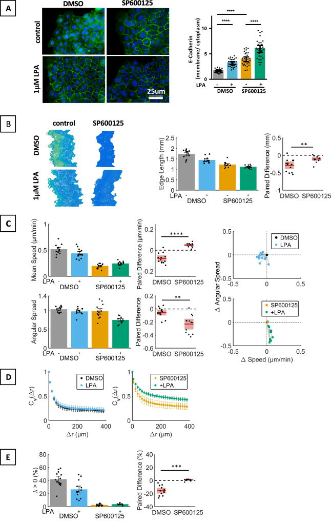
Fig. 5. Effect of inhibition of JNK signaling with SP600125 on LPA-mediated changes of the morphological and migratory phenotype of MCF10CA1a cells.
MCF10CA1a cells were starved in serum reduced medium (0.1 % horse serum) starting 3 h before stimulation with LPA (1 µM). The JNK inhibitor SP600125 (10 µM) was added 30 min before stimulation of cells with LPA. A. Immunostaining of E-Cadherin and quantification of membrane and cytoplasmic signal. ANOVA / Sidak’s multiple comparison test. n = 30 cells per group, representative of 4 experiments. Scale bar applies to all images of this panel. B–E. Effect of SP600125 and LPA on edge length (Wilcoxon matched-pairs sign rank test, B), cell speed (paired t-test) and angular spread (paired t-test) (C), Cv(Δr) (D), and λ (Wilcoxon matched-pairs sign rank test, E). n= 13 experiments.

