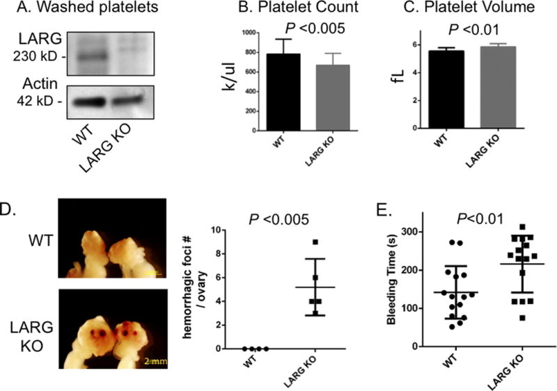
Figure 1. Platelet changes in LARG KO mice and defective hemostasis.

(A) Analysis of LARG protein expression in WT and LARG KO platelets. Actin was used as loading control. (B,C) Peripheral platelet count (B) and platelet volume (C) of WT and LARG KO littermates. Data are presented as mean + SD of 10-30 mice per group. fL indicates femtoliter. (D) Representative images of gross appearance of ovaries of 8-week-old WT and LARG KO littermates with number of hemorrhagic foci/ovary quantified in graph (WT n=4; LARG KO n=5) (E) Tail bleeding times of WT and LARG KO littermates. Each symbol represents 1 mouse (n=15/genotype).
