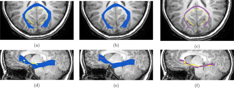
Figure 10. Registration examples.

Left image in each row shows the moving bundle (in yellow) and the reference bundle (in blue). Middle image in each line shows the two bundles registered. Right image in each line shows the moving bundle before registration (in yellow) and after registration (in purple). Top row shows an example of callosum forceps major fiber bundle registered between a 8.5 year old female and a 4 months old female. Bottom row shows an example of the left inferior fronto-occipital fasciculus (IFOF) registered between an 8.5 year old female and a 21 days old female. In both rows the background is the T1-image of the reference 8.5 year old child, axial plane on the top row and sagittal plane on the bottom row.
