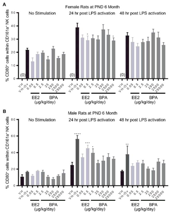
Figure 8. Percentage of CD80+ cells within the NK cell population post LPS activation from by treatment group and sex.
Female (A) and male (B) rats were administrated vehicle (VH, 0.3% aqueous carboxymethylcellulose), BPA or estrogen ethinyl estradiol (EE2) at the indicated dose level by oral gavage daily and sacrificed at postnatal day (PND) 6 months. Splenocytes were isolated and treated with LPS for up to 48 h. The percentage of CD80+ cells within CD161a+ NK cells was quantified by flow cytometry. Results are presented as mean ± SE. n = 3–10 rats/treatment group/sex. * p < 0.05, ** p < 0.01, *** p < 0.001, **** p < 0.0001 when compared to respective vehicle control (VH-Ov for female rats at PND 6 month) by a two way ANOVA with Dunnett’s posttest. For more details about VH-Ov, please see the Statistical Analysis section in the Materials and Methods.

