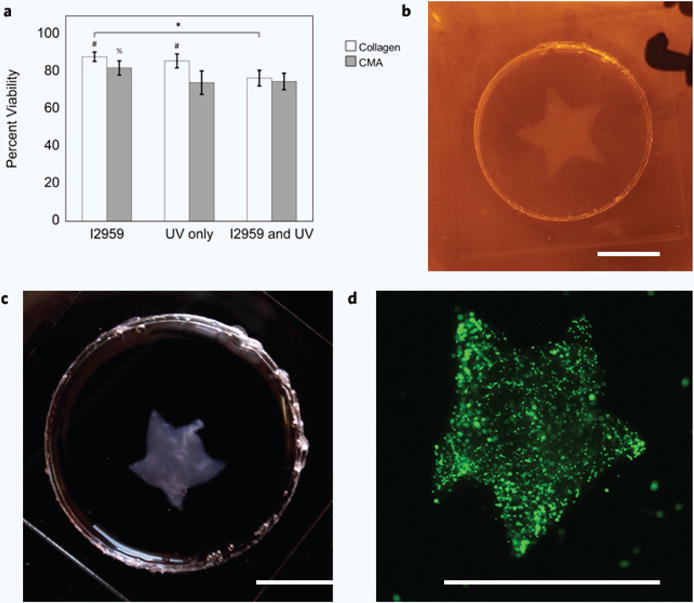
Figure 5. Cytoxicity and Free-Form Fabrication of hMSC-Encapsulated Hydrogels (a–d).

(a) Cell viability was the highest in collagen hydrogels containing I2959 (~88%), and the lowest in CMA hydrogels with Irgacure and exposed to UV (~75%). Percent cell viability decreased in each condition; hydrogels containing Irgacure had the highest percent viability, and hydrogels containing Irgacure and exposed to UV had the lowest percent viability. These differences were significant when comparing between each collagen hydrogel condition (*p < 0.05); however, CMA hydrogels containing Irgacure and were not exposed to UV had a statistically higher percent viability compared to the other CMA conditions (%p < 0.0001). Cell viability in collagen hydrogels was statistically higher compared to CMA hydrogels, except for the collagen and CMA conditions that contained Irgacure and were exposed to UV (#p < 0.0001). (b) Photopatterned, cell-laden hydrogel immediately after cold-melting. (c) Photopatterned, cell-laden hydrogel 24 hours after cell culture. (d) Calcein-AM stained hMSC in photopatterned CMA hydrogel. All photopatterned, cell-laden hydrogels demonstrated good pattern fidelity as seen in the previous figure (Fig. 4). Scale bar = 5 mm (b–d). Error bars are ± s.d.
