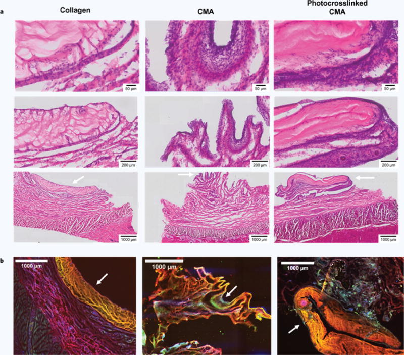
Figure 6. Histology and Immunostaining of Collagen, CMA, and Photocrosslinked CMA Implants 1 Week after Subcutaneous Implantation (a,b).

White arrows indicate scaffolds, and all histology images are oriented such that the epidermis is at the bottom of the image and the scaffold is at the top. (a) H&E stained sections at the 1-week timepoint of collagen, CMA, and photocrosslinked CMA scaffolds and tissue surrounding the implant site. All samples showed signs of an inflammatory response; the response in the tissue surrounding the collagen implant was more acute compared to that elicited by the CMA and photocrosslinked CMA samples. Cell densities in the CMA and photocrosslinked CMA sections were much higher. Signs of an inflammatory response are seen as cell immigration to the area surrounding the scaffold, with some signs of cell infiltration into the scaffold. (b) Immunostaining for rat collagen (red), bovine collagen (yellow), and cell nuclei via DAPI labeling (blue). The rat collagen antibody stained collagen non-specifically; therefore, bovine collagen is seen as yellow in the overlay instead of green. Portions of the collagen and CMA scaffolds were clearly evident in each of the sections (white arrows). DAPI labeling was significant, labeling the immediate area around the scaffold. There was also a high cell density evident in the area surrounding the scaffold, emulating the cell densities seen in H&E staining.
