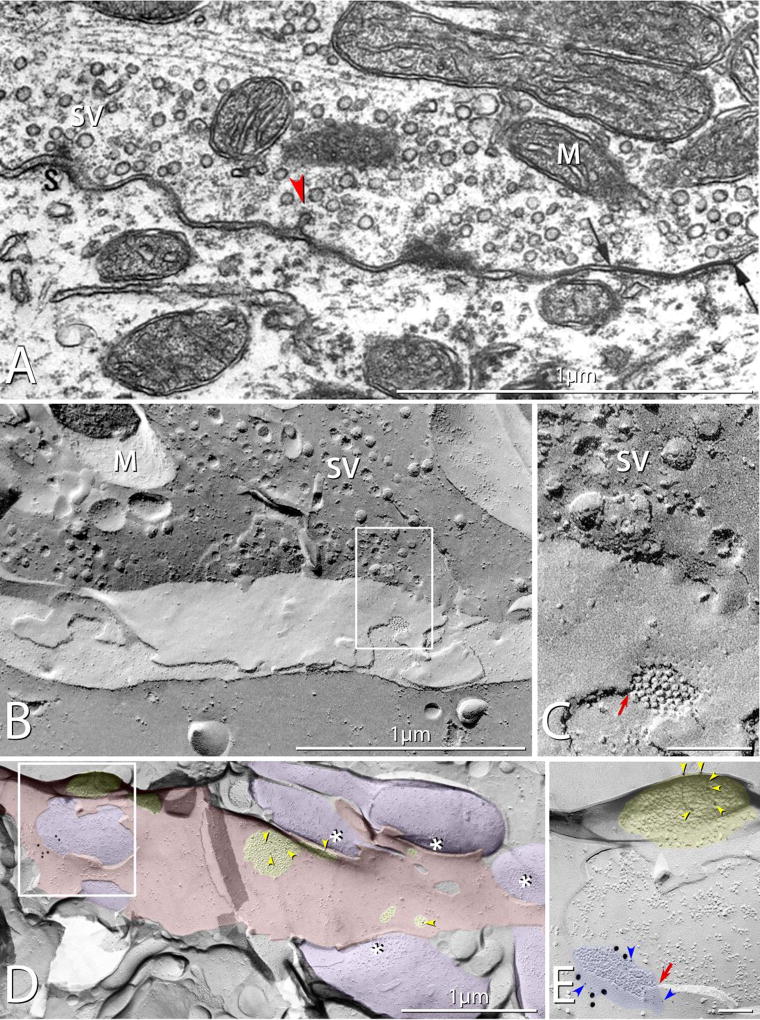
Fig. 2.
Mixed synapses in rat. (A) Thin-section TEM image of a mixed synapse in rat lateral vestibular nucleus, showing axon terminal filled with round synaptic vesicles and displaying a gap junction (arrows). Also evident is a synaptic active zone (S) with attached synaptic vesicles (SV) A nearby coated pit (red arrowhead) provides evidence for prior exocytosis and ongoing endocytosis / membrane recycling. M, mitochondrion. (Modified from [8], with permission). (B) Correlative freeze-fracture image of cross-fractured axon terminal in lumbosacral adult rat spinal cord. The axon terminal is filled with synaptic vesicles (SV), and exhibits a gap junction (box) shared with a motoneuron. (C) Higher magnification of boxed area in B, showing the gap junction, with diagnostic narrowing of the extracellular space (red arrow) at step from axonal E-face to motor neuron P-face, and regular arrays of P-face particles and E-face pits. (D,E) Low- and high-magnification images of multiple synaptic terminals onto unidentified spiny dendrite in stratum oriens in CA3 region of adult rat hippocampus. Active zones on some axon terminals are indicated by asterisks; PSDs (yellow overlays) contain immunogold-labeled NMDA receptors. (E) A higher magnification image of boxed area in D, showing a gap junction linking one of the axon terminals (purple overlays) to the dendrite (red overlay). The gap junction is heavily labeled for Cx36 (21 6-nm gold beads [blue arrowheads] and six 18-nm gold beads), and the nearby glutamate receptor-containing PSD, recognized as clustered 9–10 nm E-face particles (yellow overlay), is immunogold labeled for NMDA receptors (12-nm gold beads; yellow arrowheads). Note that the axon terminal shares the gap junction and the PSD with the overlying E-face of the small-diameter dendrite. (Modified from [105])

