Fig. 5.
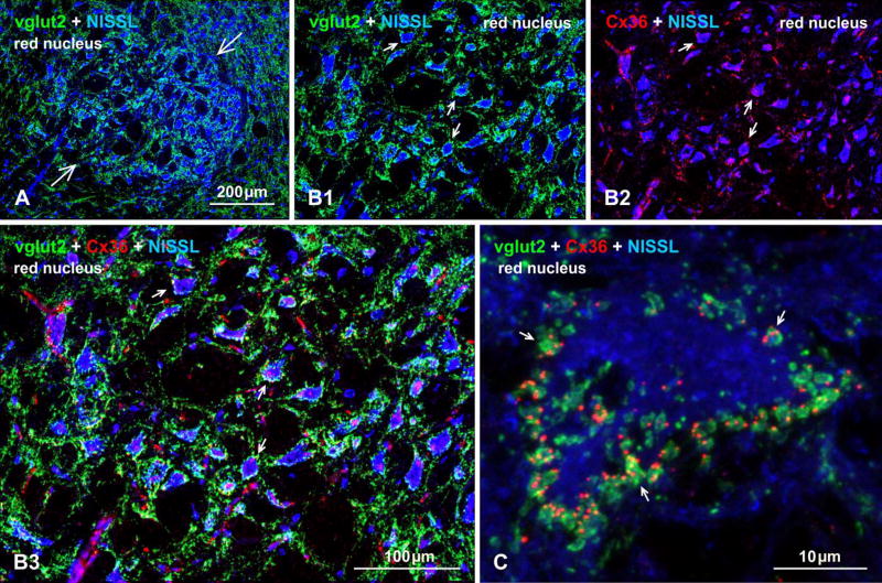
Immunofluorescence evidence for mixed synapses formed by axon terminals and neurons in the red nucleus of adult mouse brain. (A) Image of the red nucleus (arrows) with its distinctive large neurons counterstained with blue fluorescence Nissl, and showing a high density of immunolabeling for vglut2 within the nucleus. (B) Higher magnification of the red nucleus, showing vglut2-positive nerve terminals heavily concentrated around blue Nissl-stained neuronal somata and initial dendrites (B1, arrows) and, in the same field, immunolabeling of Cx36 associated with the same Nissl-stained neurons (B2, arrows) and often displaying overlap with labelling of vglut2, as shown in three-color overlay (B3, arrows). (C) Confocal image of a single large red nucleus neuron (blue fluorescence Nissl counterstained), showing the periphery of the neuronal somata heavily invested with prominent vglut2-positive terminals that frequently display co-localization with fine punctate labelling for Cx36 (arrows). Immunofluorescence labelling of Cx36 was absent in the red nucleus of Cx36 null mice (not shown).

