Figure 5.
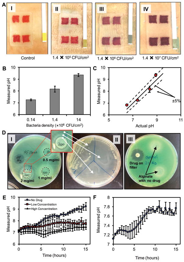
Ex vivo detection of bacterial infections and evaluation of the antibacterial efficiency of drug-eluting gel using bacteria inhibition assay. A-i–iv) Colorimetric detection of bacterial infection in pig skins. Skins were inoculated at initial densities of 1.4 × 10⁵, 1.4 × 10⁶, and 1.4 × 10⁷ CFU cm−2. After 12 h, GelDerm was placed on samples and images were taken to quantify the pH of the infected skins. B) pH values of infected skins measured by smartphone. C) Comparison of the GelDerm readings with commercially available pH strips. D) Drug-eluting hydrogel disks containing 0.5, 1, and 2 mg mL−1 of gentamicin on agar sheets along with a paper disk containing 1 mg mL−1 of the same drug as positive control condition. P. aeruginosa was cultured on the sheets to observe the effect of the hydrogels on the bacteria. D-i) A clear ring was observable around the 200 mg mL−1 hydrogel. D-ii) Samples from three different areas of the ring surrounding the 200 mg mL−1 hydrogel were cultured in a different Petri dish to determine whether there is bacterial growth in those areas. D-iii) Hydrogels without any drug content were placed in a separate Petri dish as negative control condition. E) The pH change of pig skin samples inoculated with P. aeruginosa during a 15 h period with three different conditions for drug concentration—0 (no drug), 0.5 mg mL−1 (low concentration), and 2 mg mL−1 (high concentration). F) The pH change of pig skin samples infected with 1:1 mixture of P. aeruginosa and S. aureus. Error bars are SD (n = 3).

