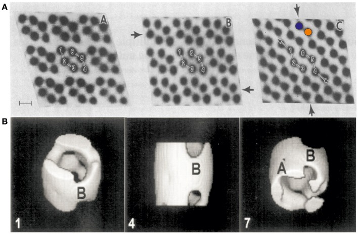
Figure 1.
First electron micrographs of VDAC from the early 1980s. (A) Electron-microscopic investigations of ncVDAC arranged in isolated native membrane vesicles show three two-dimensional molecular arrays: two slightly different hexameric arrangements of VDAC pores around a two-fold symmetry axis (right and left) and another arrangement where dimeric VDAC pores form chain-like superstructures (colored circles mark two independent monomers) (Mannella et al., 1983). (B) A single particle analysis of small membrane arrays yielded the first 3D representation of VDAC at a resolution of ~2 nm (Guo et al., 1995). Figures reproduced with permission.

