Figure 4.

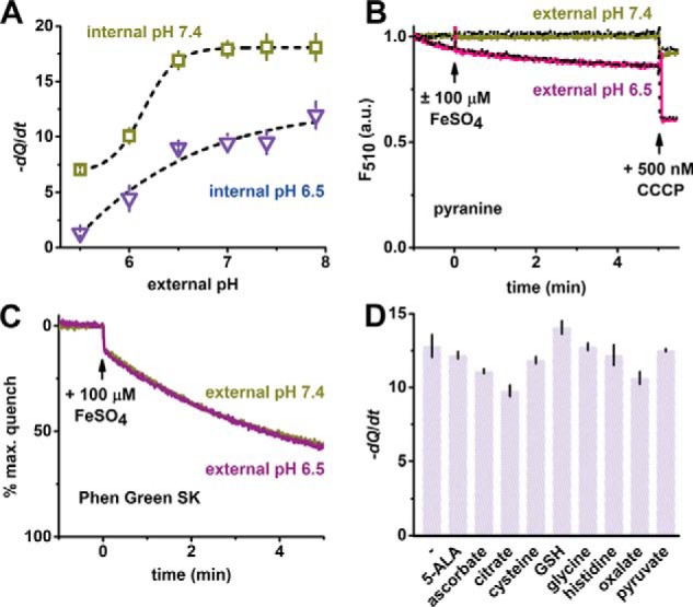
Modulation of TMfrn1 iron transport by pH and ligands. A, pH dependence of TMfrn1 iron transport. Points depict initial quenching rates after iron addition. Black lines are sigmoidal fits of quenching rates at internal pH 7.4 and 6.5. B, TMfrn1 iron transport does not alter intraliposome pH. Measurement of pH-dependent pyranine fluorescence in the presence or absence of iron. Solid colored lines show addition of iron to TMfrn1 proteoliposomes; dotted black lines lack iron. Liposomes contain 50 μm pyranine and contents are weakly buffered with 2 mm MOPS, pH 7.4, 0.5 mm EDTA, 140 mm KCl. Carbonyl cyanide m-chlorophenyl hydrazine (CCCP) was added as indicated to dissipate proton gradient. C, weakly buffered TMfrn1 proteoliposomes retain iron uptake. Liposome internal contents are similar to A except PGSK (instead of pyranine) was used, and no EDTA was added. D, labile iron pool candidates have negligible effect on TMfrn1 iron transport. A 100 μm concentration of each iron ligand was added to the TMfrn1 proteoliposome suspension followed by 10 μm iron, and PGSK fluorescence quenching was monitored. Plotted are initial PGSK quenching rates. Assays were performed with internal pH 7.4 TMfrn1 proteoliposomes diluted into external pH 6.5 buffer. 5-ALA, 5-aminolevulinic acid; GSH, glutathione; a.u., arbitrary units. In A and D, error bars depict S.D. of triplicates.
