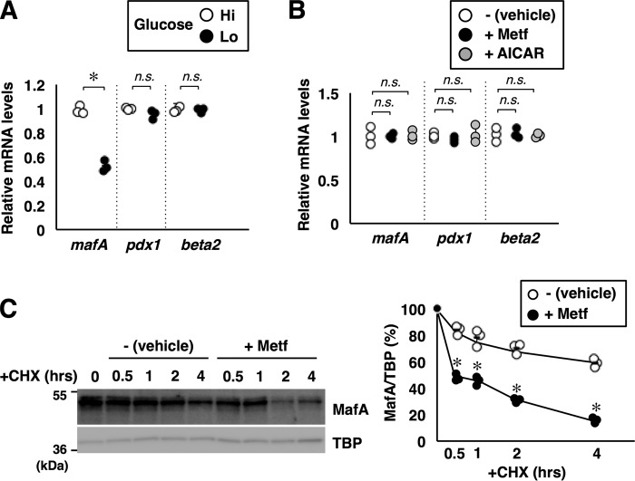
Figure 4.
AMPK promotes MafA protein degradation. A, total RNA isolated from MIN6 cells grown under high-glucose (Hi, 20 mm) or low-glucose (Lo, 2 mm) conditions for 16 h were analyzed by quantitative RT-PCR using specific primers. Data are mean ± S.E. of three independent experiments. *, p < 0.05; n.s., not significant. B, MIN6 cells grown in high-glucose medium were treated with metformin (Metf, 500 μm) or AICAR (200 μm) for 16 h. Total RNA was then isolated and subjected to quantitative RT-PCR analysis. Data are mean ± S.E. of three independent experiments. C, degradation rates of endogenous MafA protein. MIN6 cells treated with or without metformin (500 μm) for 16 h were incubated with cycloheximide (CHX, 10 μg/ml) for the indicated periods, and cell extracts were analyzed by immunoblotting. TBP was used as a loading control because TBP seems to be a long-lived protein in MIN6 cells. Signal intensities of MafA relative to TBP are shown in the right panel. Data represent the results of three independent experiments. *, p < 0.05; **, p < 0.01 (Student's t test).

