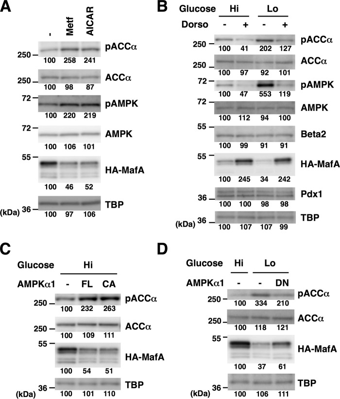
Figure 5.
Glucose and AMPK regulate exogenously expressed MafA protein. A, an expression plasmid containing HA-MafA under the control of the EF1α promoter was transfected into MIN6 cells, and the cells were treated with metformin (Metf, 500 μm) or AICAR (200 μm) for 16 h in high-glucose medium. The cell extracts were analyzed by immunoblotting using the indicated antibodies. B, MIN6 cells transfected with the HA-MafA expression plasmid were grown in high-glucose (Hi, 20 mm) or low-glucose (Lo, 2 mm) medium and treated with dorsomorphin (Dorso, 10 μm) for 16 h. Cell extracts were then subjected to immunoblot analysis. C, the HA-MafA expression plasmid was co-transfected into MIN6 cells with an expression plasmid for FL or CA AMPKα1. Cell extracts were analyzed by immunoblotting. D, MIN6 cells were transfected with expression plasmids for HA-MafA and a DN form of AMPKα1, and the cells were incubated in medium containing the indicated concentrations of glucose for 16 h. Whole-cell extracts were subjected to immunoblot analysis. The data in A–D are representative of two independent biological experiments. The intensity of the bands was quantified using ImageJ software, and the relative amounts (averages of two independent experiments) are indicated below the bands.

