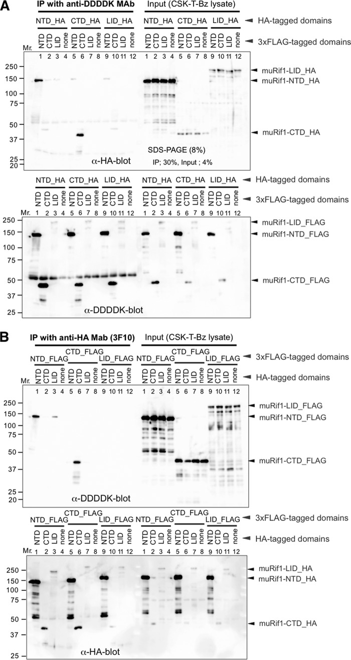
Figure 2.
Self-association of muRif1-NTD and of muRif1-CTD. Various portions of muRif1 were fused to either HA tag or 3xFLAG tag, and combinations of an HA-tagged and a FLAG-tagged polypeptide were transiently expressed in 293T cells as shown. One portion of cell lysates was subjected to immunoprecipitation (IP) with anti-DDDDK antibody (A) and the other portion with anti-HA antibody (B). Precipitated (left) and input (right) polypeptides were detected by Western blotting with anti-HA antibody (upper) or with anti-DDDDK antibody (lower). muRif1-LID showed weak signals, because electrotransfer efficiency of muRif1-LID from polyacrylamide gel to PVDF membrane was low, probably due to its very acidic property (calculated pI = 4.64).

