
Figure 2.
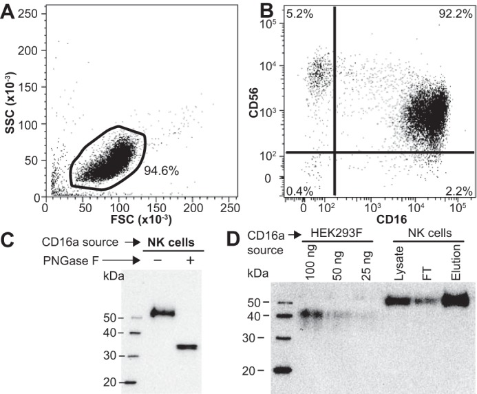
Representative NK cell isolation and CD16a purification. A, light scattering; B, double-antibody staining of negatively-selected NK cells. Isotype and negative-staining controls are shown in Fig. S1. C, anti-CD16 Western blot of PNGase F-digested CD16a shows an increase in mobility following N-glycan removal. D, anti-CD16 Western blot of CD16a purification from an NK cell lysate compared with recombinant CD16a truncated at the transmembrane domain (HEK293F).