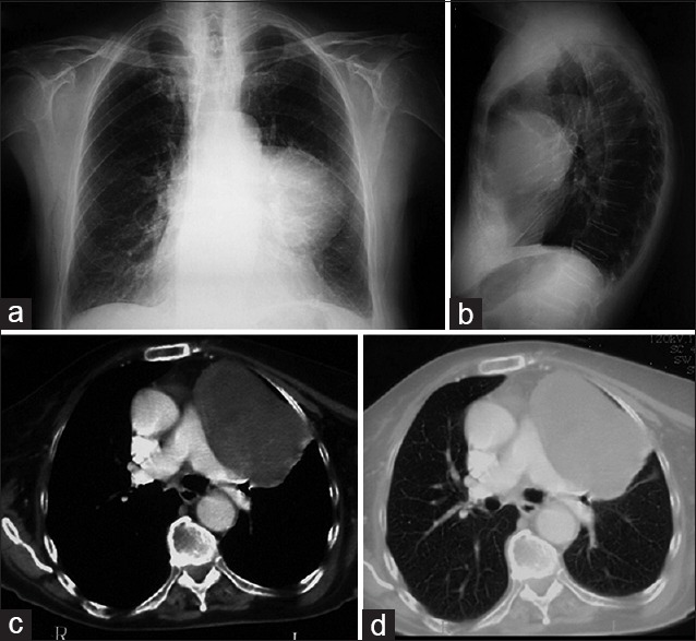
Figure 7.

A 78-year-old woman with fever underwent a chest X-ray that showed a large opacity localized in the retro-sternal space (a and b). Enhanced CT scans demonstrated a well-defined round lesion (10 x 9 cm), heterogeneous, without signs of infiltration of mediastinum which occupies part of the thymic lodge extending in the pleural cavity (c and d). Left lateral thoracotomy with wedge resection was performed; the large mass was pedunculated, growing up from the lingular visceral pleura.
