Figure 1.

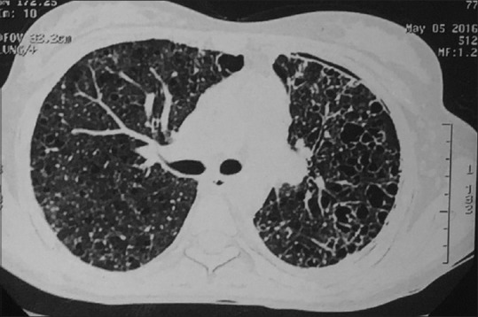
High-resolution computed tomography thorax at the level of right main bronchus showing bilateral thin-walled cystic lesions and left-sided pneumothorax

High-resolution computed tomography thorax at the level of right main bronchus showing bilateral thin-walled cystic lesions and left-sided pneumothorax