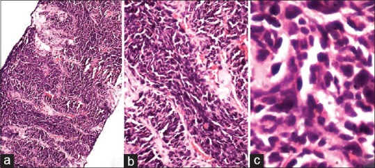
Figure 2.

(a-c) Section show infiltrating lesion comprised of sheets and trabeculae of polygonal cells. These cells have high N:C ratio with scant cytoplasm. Nuclei show evidence moulding and spindling with inconspicuous nucleoli (H and E; [a] ×40, [b] ×100, [c] ×400)
