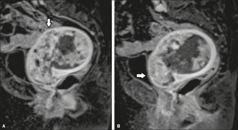
Figure 2.
MRI scan from an outside facility showing the pelvis of a 63-year-old female with endometrial cancer. Gadolinium-enhanced fat-suppressed sagittal T1-weighted images show cancer invading more than half of the myometrium (A) and the cervical stroma (B). The initial report described an endometrial tumor invading less than half of the myometrium. Subsequent histopathology confirmed the findings of the second-opinion interpretation.

