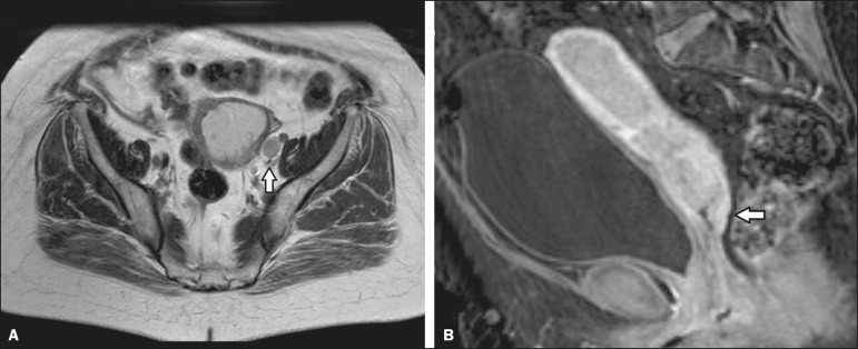
Figure 3.
Axial T2-weighted image (A) and gadolinium-enhanced fat-suppressed sagittal T1-weighted image (B) from an outside facility. Neither the enlargement of the left external iliac lymph node nor the invasion of the upper posterior third of the vagina was reported in the initial interpretation of the MRI scans of this patient with endometrial cancer invading more than half of the myometrium and the cervical stroma. Although those findings led to preoperative upstaging, the decision made by the multidisciplinary board was that radiotherapy would have been the first approach to treatment in either case. Therefore, the second-opinion report did not affect the management in this particular case.

