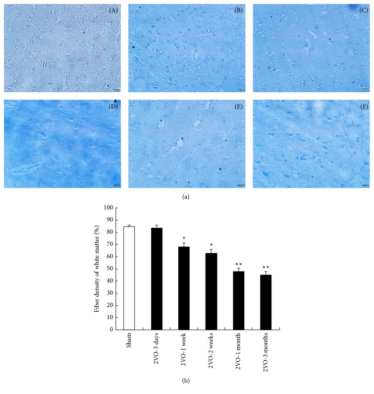
Figure 1.
Fiber densities being stained with Luxol fast blue in the deep white matter of the CCH rats (a) and quantitative fiber densities (b). From direct microscopical observations, LFB staining was slightly affected at 3 days (B), 1 week (C), and 2 weeks (D). At 1 and 3 months after 2VO (E, F), the fiber density was significantly decreased compared with that of the sham control (A). (∗p < 0.05, ∗∗p < 0.01, compared with the brain of sham control animals; n = 5 in each group, LFB 200x).

