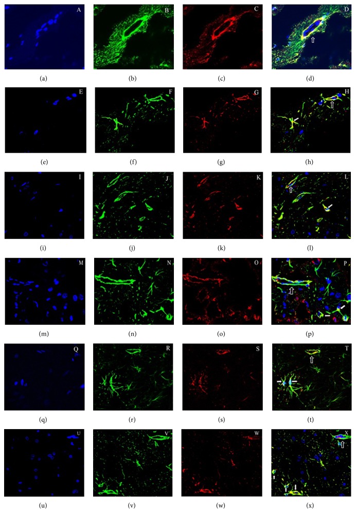
Figure 5.
Immunofluorescence colocalization for GFAP and AQP4 in the brain. The blue nucleus in the brain was DAPI labelled. AQP4 immunohistochemistry labelling in astrocytes was red. GFAP positive astrocytes in the brain were labelled green. The coexpression of GFAP and AQP4 within the same cell is yellow (merged). Sham group (a, b, c, and d), 2VO-3 days' group (e, f, g, and h), 2VO-1-week group (I, j, k, and l), 2VO-2 weeks' group (m, n, o, and p), 2VO-1-month group (q, r, s, and t), and 2VO-3 months' group (u, v, w, and x), respectively. AQP4 was obvious on astrocyte end-feet membranes in the sham group with yellow colour as symbol, but the yellow colour had been changing from end-feet membranes to astrocytic body since 3 days after 2VO, and the alteration had been persisting even 3 months after 2VO (IF 600x).

