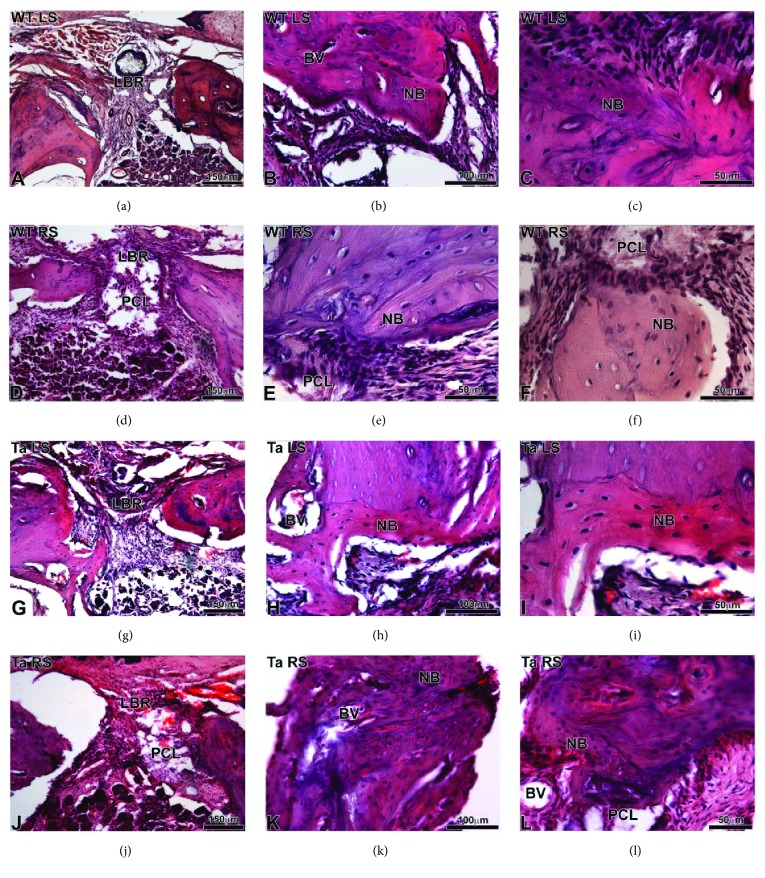
Figure 7.
Histological sections of WT (a–f) and Ta (g–l) maxillary bone stained with hematoxylin-eosin after 30 days of implantation. (a–c) and (g–i) corresponded to the lesion on the left side without scaffold. (d–f) and (j–l) corresponded to the lesion on the right side with bifunctionalized BMP-2/Ibuprofen scaffold. BV: blood vessel, LBR: lesion with bone regeneration, NB: neoformed bone, and PCL: bifunctionalized scaffold.

