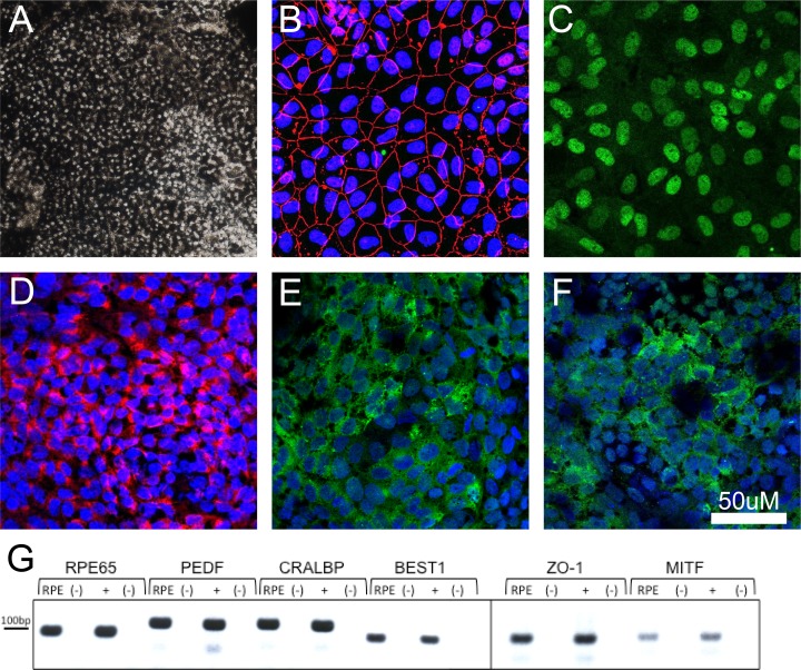
Figure 1.
Characterization of rhesus monkey iPS-derived RPE cells. (A) Image captured from a confluent and mature plate of RPE cells. (B–F) Images of RPE cells stained with the immunohistochemical markers ZO-1 (B), MITF (C), PMEL17 (D), CRALBP (E), and RPE65 (F). (G) Expression of RPE-associated genes in iPS-derived RPE as demonstrated through RT-PCR. Headings for (G): RPE, Rhesus iPS-RPE cells; (+), rhesus primary RPE tissue; (-), no-RT controls for each reaction.

