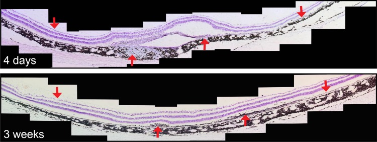
Figure 5.
Image montage of cresyl violet–stained retina sectioned horizontally through the location of the RPE cell transplant. Upward red arrows indicate the location of mononuclear cell infiltration and fibrotic scarring; downward arrows indicate loci of normal RPE and neural retina outside the transplant area.

