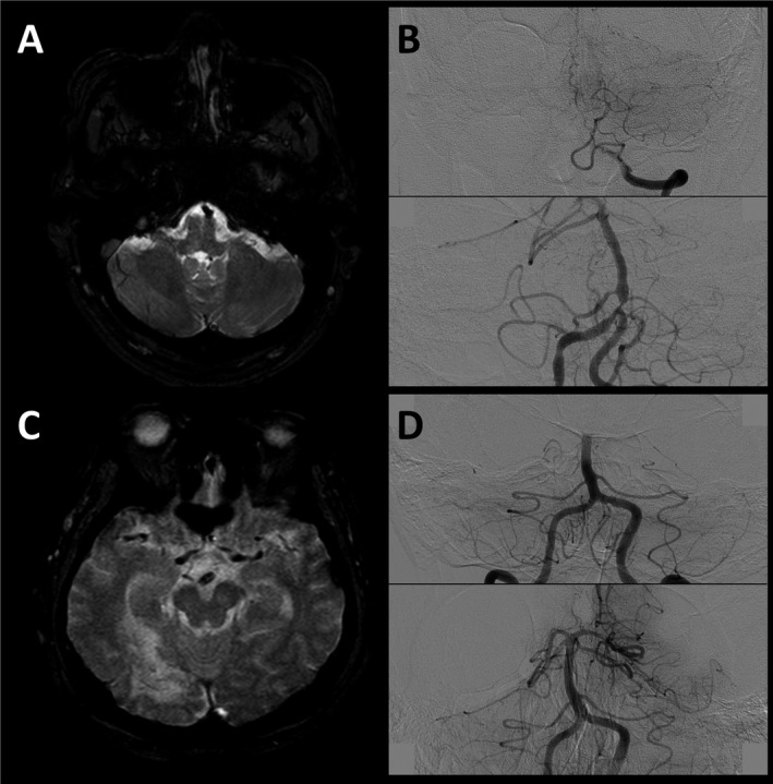
Figure 2.

Representative cases. A 60‐year‐old male patient with initial NIH Stroke‐Scale (NIHSS) score of 18. In axial gradient‐echo images (GRE) of initial MRI, a clot in the proximal basilar segment is observed (panel A). Angiography reveals a basilar artery (BA) occlusion at the vertebra‐basilar junction level (panel B, upper). Immediate follow‐up angiography after achieving a successful reperfusion by endovascular treatment revealed a tight residual stenosis in the proximal BA, suggesting an in situ atherosclerotic stenosis mechanism (panel B, lower). In GRE images of an 85‐year‐old female patient with initial NIHSS score of 21, clot sign was detected in the distal basilar segment (panel C). BA was occluded at the middle segment (panel D, upper), and no residual stenosis was observed after a complete reperfusion, suggesting an embolic mechanism (panel D, lower).
