Figure 1.

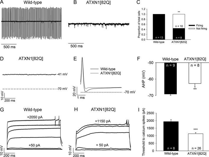
ATXN1[82Q] Purkinje neurons display both an absence of repetitive spiking and dendritic hyperexcitability. (A) Representative spiking of a wild‐type Purkinje neuron in the cell‐attached recording configuration. (B) Representative trace of a nonspiking ATXN1[82Q] Purkinje neuron in the cell‐attached recording configuration. (C) Summary of spiking and nonspiking Purkinje neurons from wild‐type and ATXN1[82Q] mice. (D) Representative trace of a nonfiring ATXN1[82Q] Purkinje neuron in the whole‐cell recording configuration. These neurons display a depolarized resting membrane potential. (E) After‐hyperpolarization (AHP) amplitude in wild‐type and ATXN1[82Q] Purkinje neurons. (F) Summary of AHP amplitudes in wild‐type and ATXN1[82Q] Purkinje neurons. (G) Representative trace of a wild‐type Purkinje neuron held at −80 mV in the presence of tetrodotoxin. Upon injection of positive current in +50 pA increments, dendritic calcium spikes are noted. (H) Representative trace of dendritic calcium spike analysis from an ATXN1[82Q] Purkinje neuron. (I) Summary of the threshold of injected current required to elicit dendritic calcium spikes in wild‐type and ATXN1[82Q] Purkinje neurons in the presence of tetrodotoxin. **P < 0.01, ***P < 0.001, Fisher's exact test (C) or two‐sample Student's t‐test (I).
