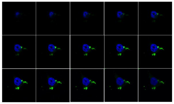
Figure 4.
Series of confocal microscopy “optical slices” of a cell treated with azide nanoparticles labeled by Click reaction with alkyne-Alexa Fluor 488 in situ. Single-cell optical slices of 0.22 μm show the cytoplasmic localization of nanoparticles Click labeled in situ (green) at different focal depths inside the cell. Cell nuclei were labeled by Hoechst staining (blue).

