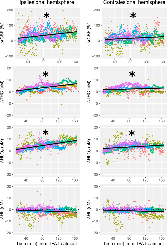
Fig. 2.
Frontal microvascular relative cerebral blood flow (rCBF), total hemoglobin concentration (THC), oxy-hemoglobin (HbO2), and deoxy-hemoglobin (Hb) changes over time are shown for the ipsilesional hemisphere (left column) and the contralesional hemisphere (right column) for all five patients (each patient in different color). (Δ) indicates a change of the optical variable from the baseline. Black line shows the fitted linear model of all the measurement points plotted. The time, x-axis, shows the time elapsed from the rtPA treatment onset. (*) indicates a statistically significant positive slope (p<0.05).

